
Project NoMBSI
(Non-lethal Method of Benthos Sampling and Identification)

NoMBSI (Non-lethal Method of Benthos Sampling and Identification) is a method, in general, comprising the same stages as have been employed in traditional methods using in biological assessment:
- sampling (follow),
- sample preservation (follow),
- taxa identification (follow),
- interpretaion of results (follow).
However, the particular stages differ in their details. Since the most harmful stages in terms of animal mortality are sample preservation and taxa identification, both of these stages have been made subject to far-reaching modifications, in that “digital preservation” and images analysis take the place of sample preservation in alcohol and taxa identification using a stereoscopic microscope.
Sampling
|
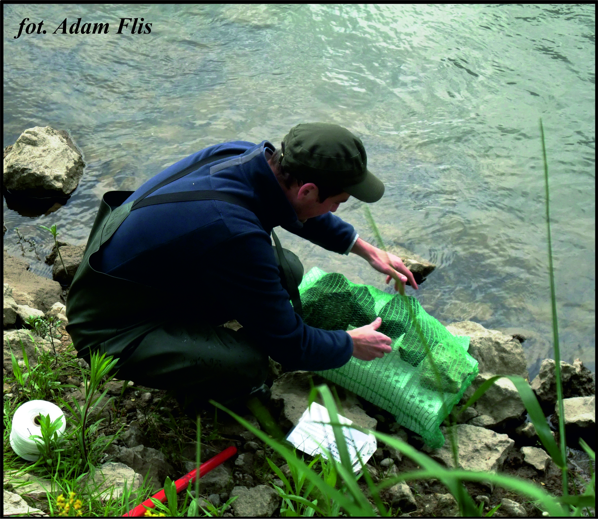
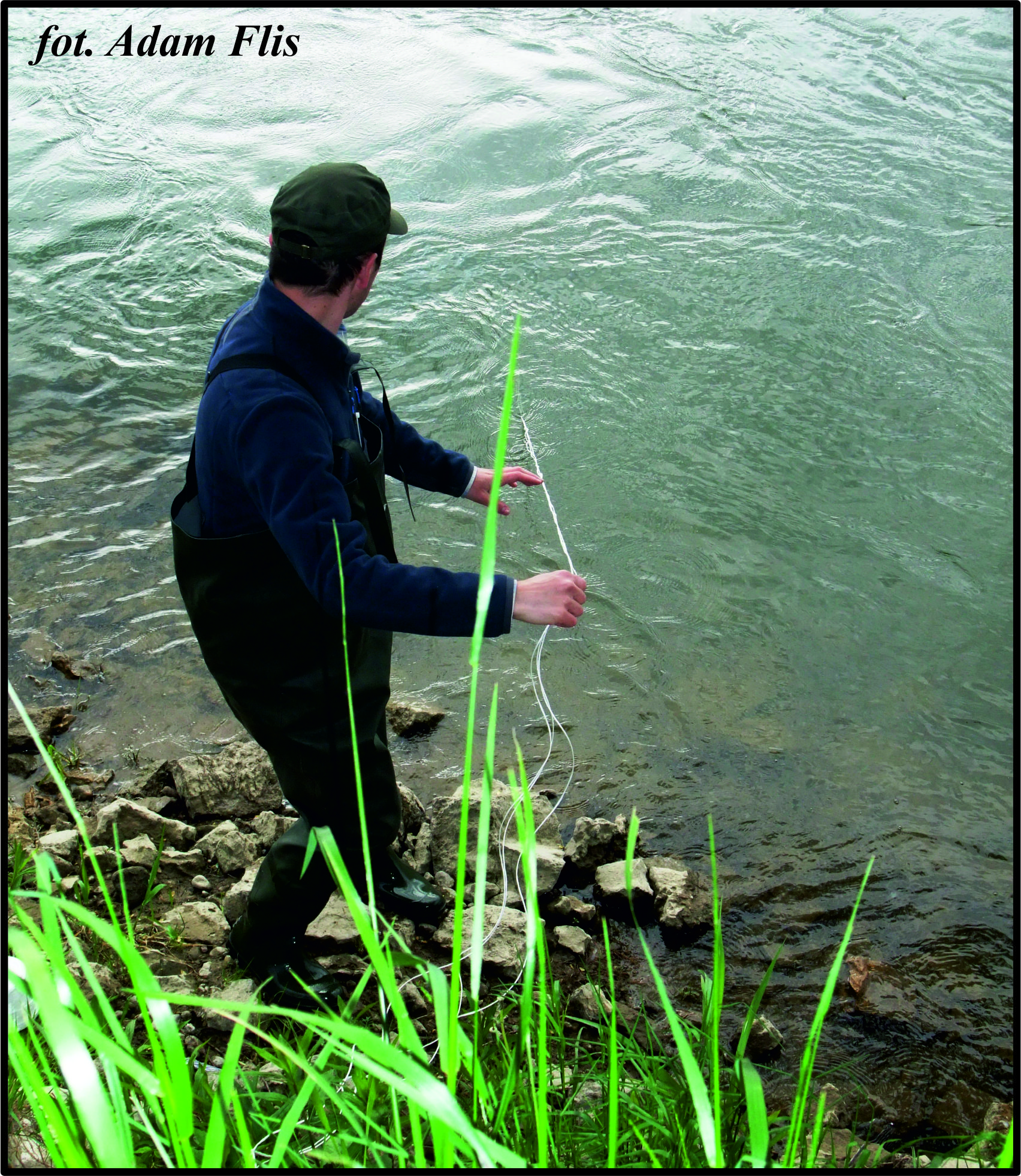
NoMBSI procedure is based on artificial substrate samplers (AS) placed in a riffle, see Images 1 and 2 below (e.g. De Pauw et al., 1986 [2]; Czerniawska-Kusza, 2004 [1]).
|
|
|---|---|
|
|
Images 1 (upper left) and 2 (upper right): Artificial substrate placement in water.
|
|
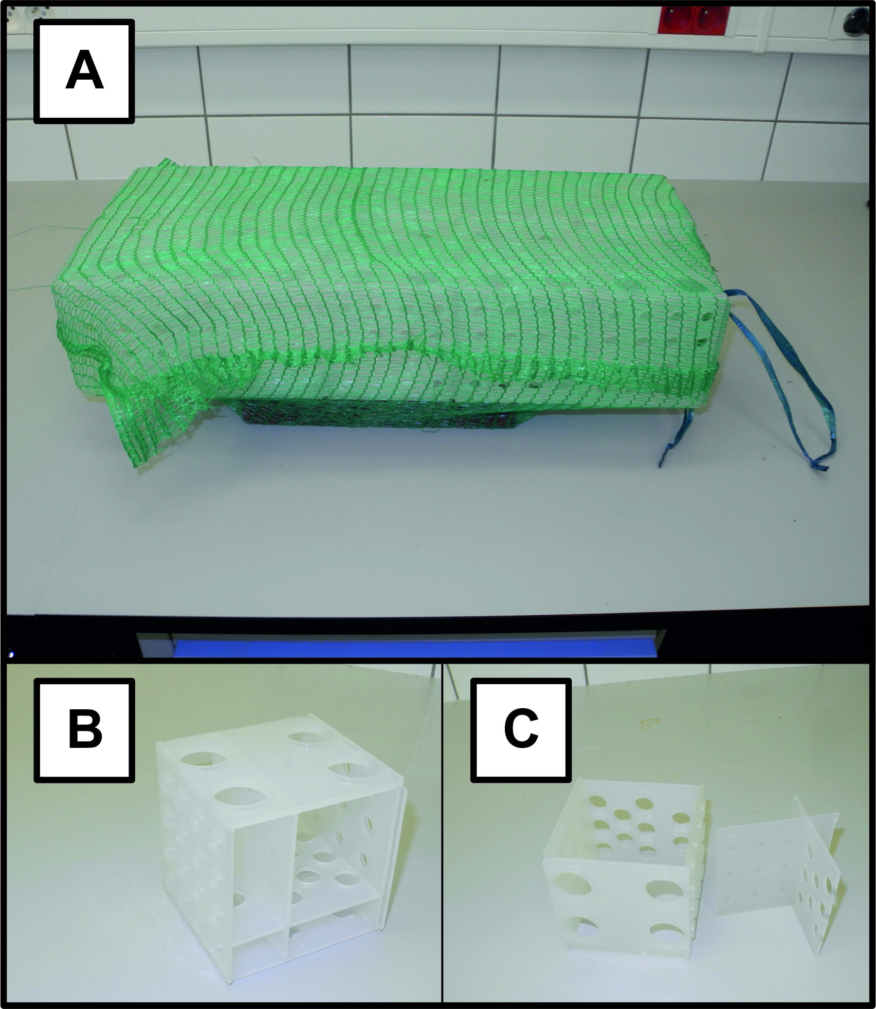
In our research the single AS is constructed out of a set of ten combined, polypropylene cubic test tube racks. The whole set is placed into a potato bag and weighted down with stones (Image 3, on left).
The time required for artificial substrate colonisation by macroinvertebrates must be taken into consideration when the length of a period when AS is left under water is determined. Six weeks seems to be most convenient time in case of rivers (e.g. De Pauw et al., 1986 [2]). |
Image 3 (upper left): Image of artificial substrate sampler (AS) uses for macroinvertebrates sampling in non-lethal NoMBSI procedure. A – a single AS setup, ready for placing in sampling site. B – a single cubic test tube rack used for AS construction. C – a single cubic test tube rack taken apart – it is really helpful during animals flushing out. |
|
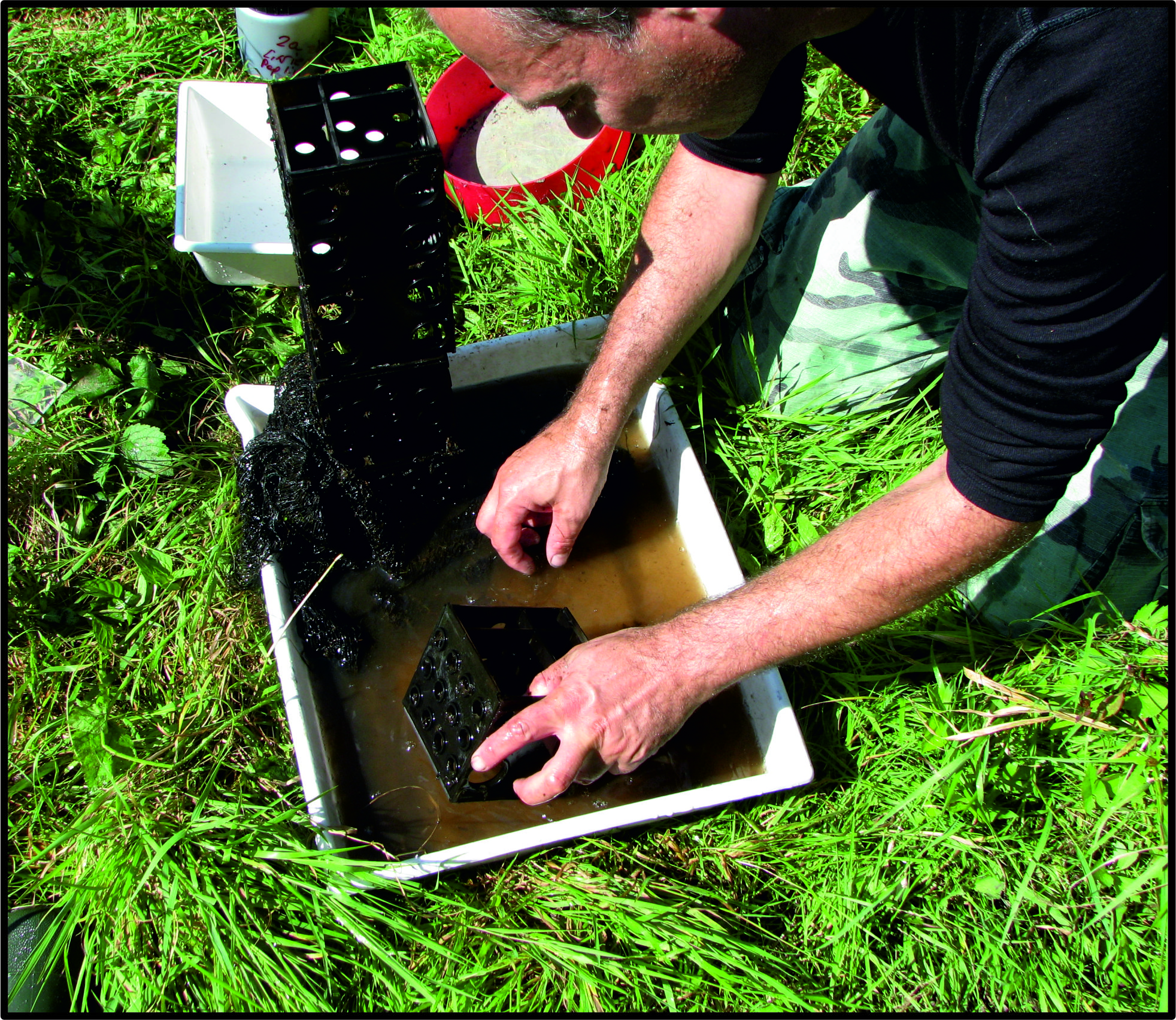
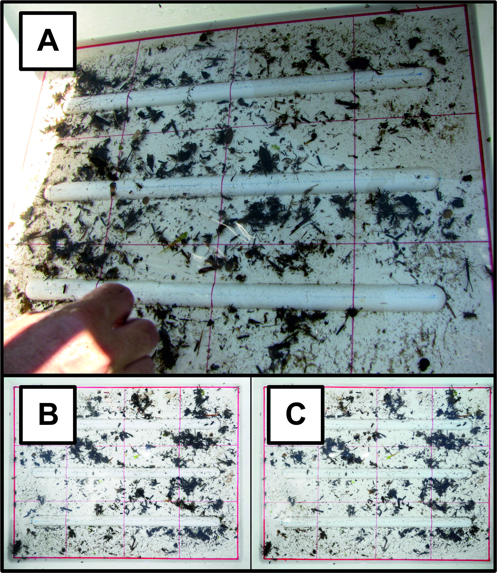
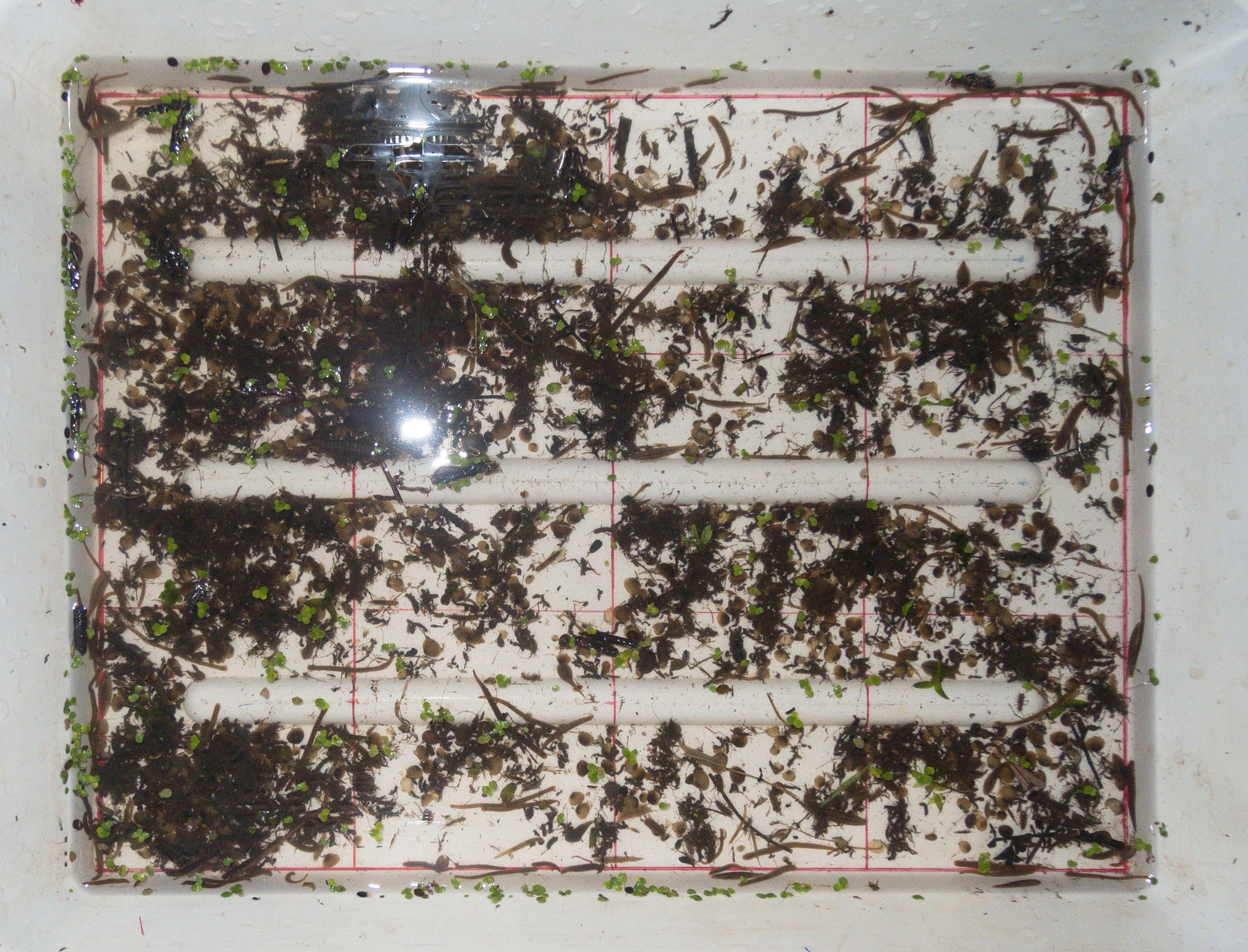
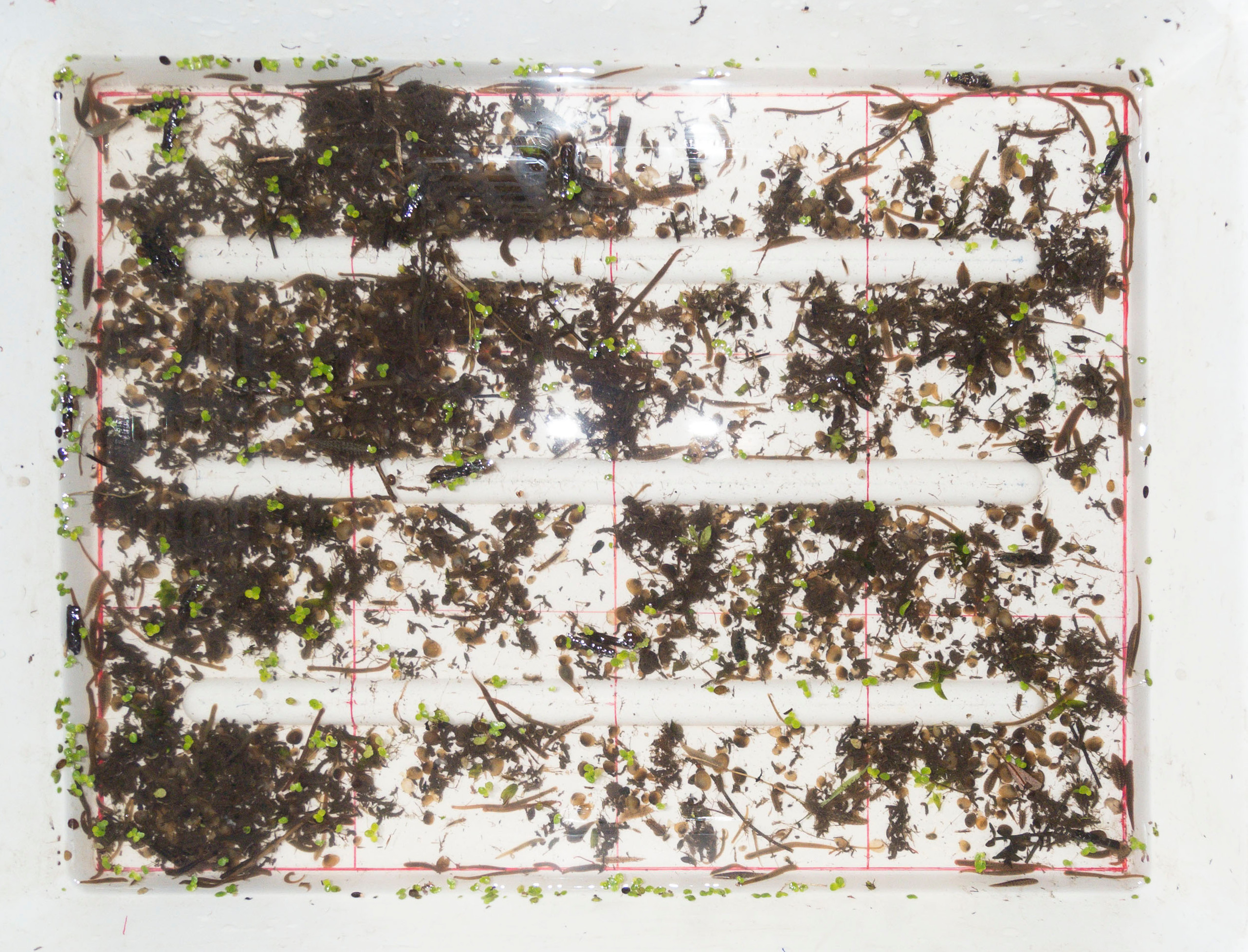
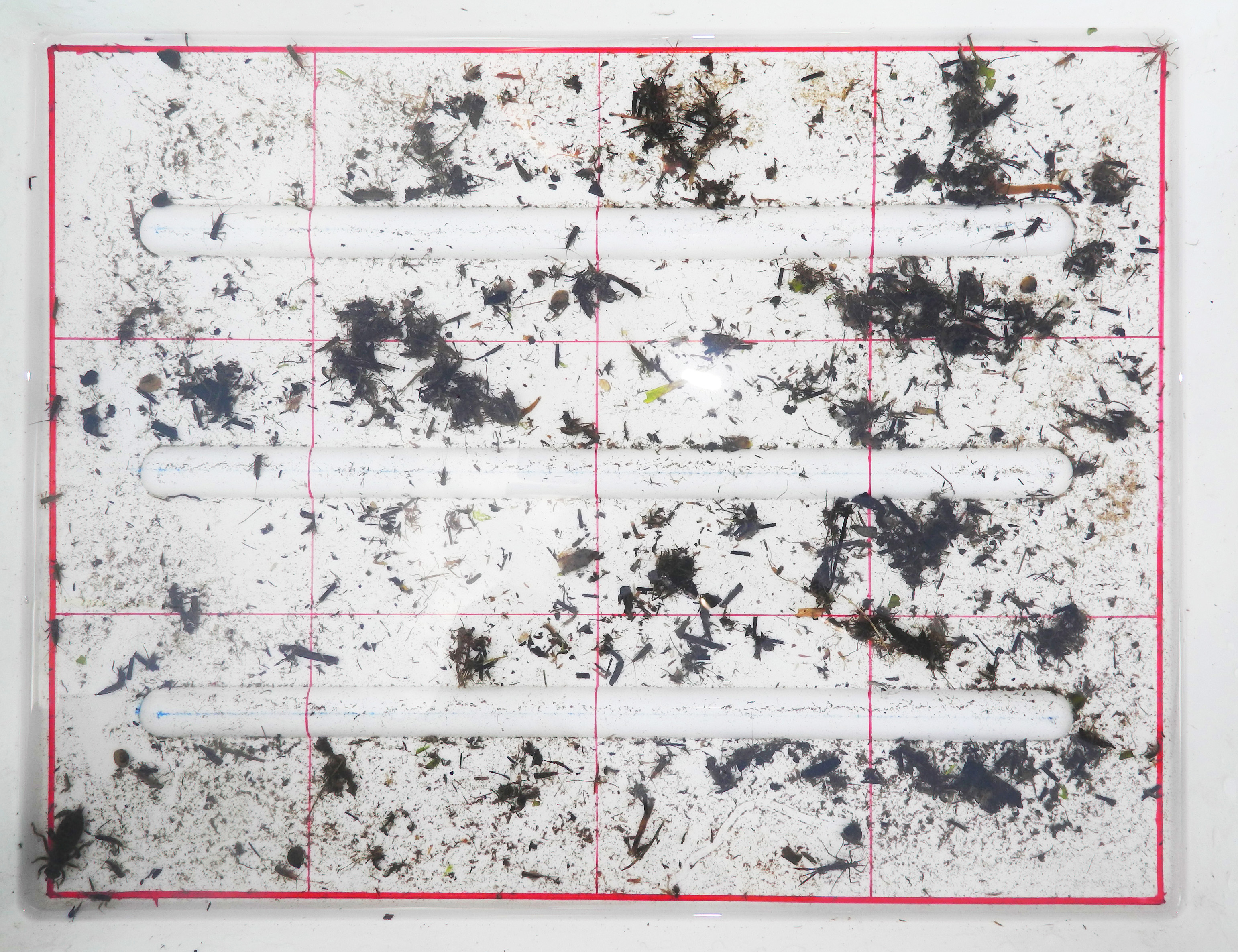
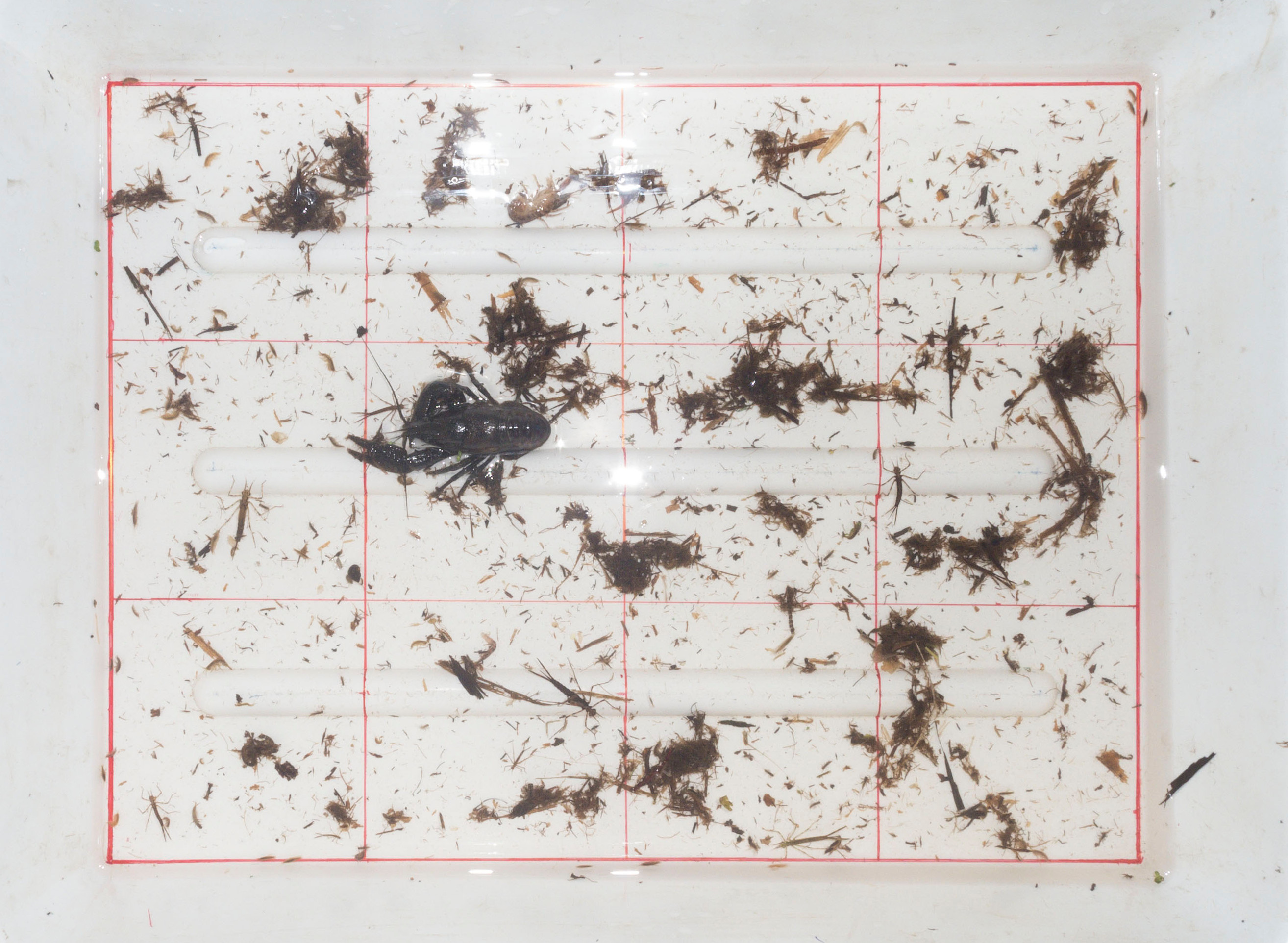
Following removal of the AS, animals present are flushed out (Image 4 below, on left), sieved (in 1 mm mesh net) and placed in a photographic tray (Image 5A below, on right). Samples should be taken in spring or late summer (or autumn), avoiding time of the year when insects are emerging and a lot of important taxa are absent in water (July). |
|
|
|
Image 4 (upper left): Flushing out of animals colonizing artificial substrate. Image 5 (upper right): Sample of macroinvertebrates placed in a photographic tray. A – image of photographic tray with prepared sample – close-up. B and C – 2D images of digitalized sample taken by left (B) and right (C) cameras of transportable photographic studio. |
(back to top of page)
Sample preservation
|
Non-lethal analysis of taxonomic composition of macroinvertebrates is the main assumption of NoMBSI. Due to this fact a whole samples of animals are digitalized in a form of three dimensional images (3D). Thereby, animals can be, in turn remove to the water at the end.
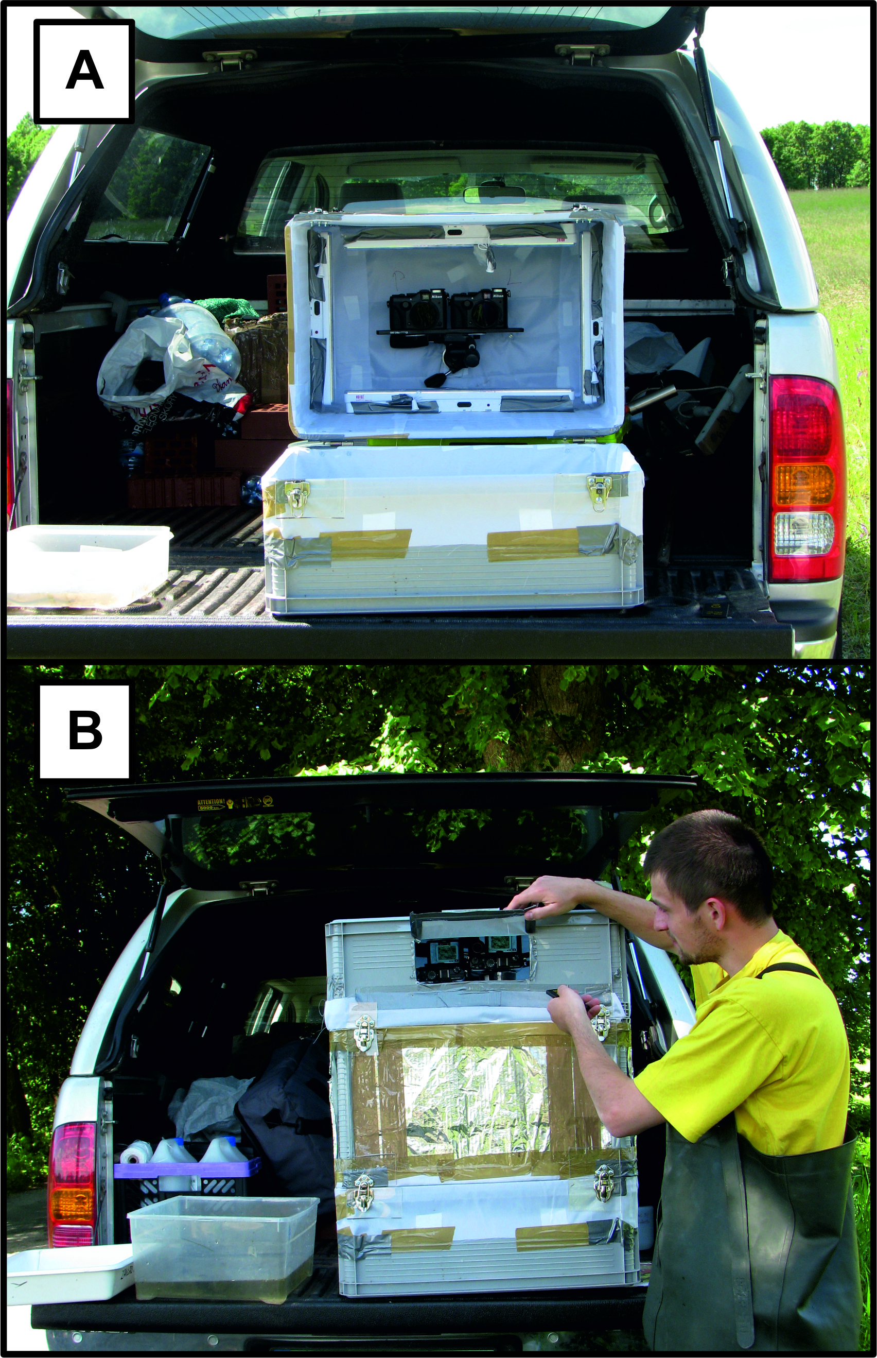
Collected samples are digitalized with the aid of transportable photographic studio – 3DS. Therefore, previously prepared photographic tray with a sample is placed on the bottom of 3DS and a sample is preserved digitally with use of two synchronized photographic cameras. 3DS is in the form of a non-transparent plastic box equipped with a place for photographic tray with a sample on the bottom, and a set of two digital photographic cameras attached to the upper cover (Image 6 A and B, on the left). |
Image 6 (upper left): Transportable photographic studio. A – a studio before placement a photographic tray with digitalized sample. A set of two digital cameras is also visible. B – taking a photo of a sample with the aid of infrared remote controle. |
|---|
(back to top of page)
Taxa identification
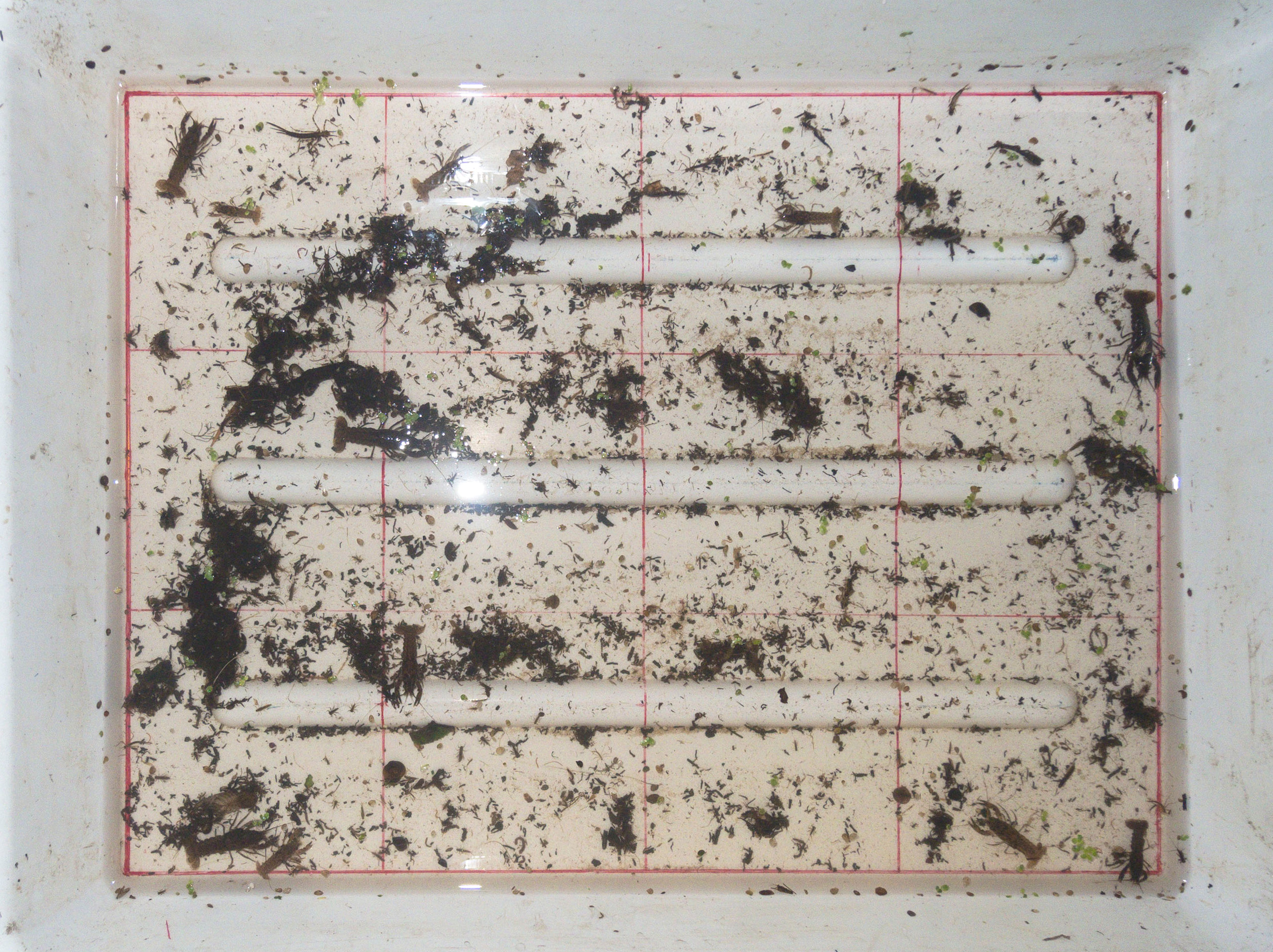
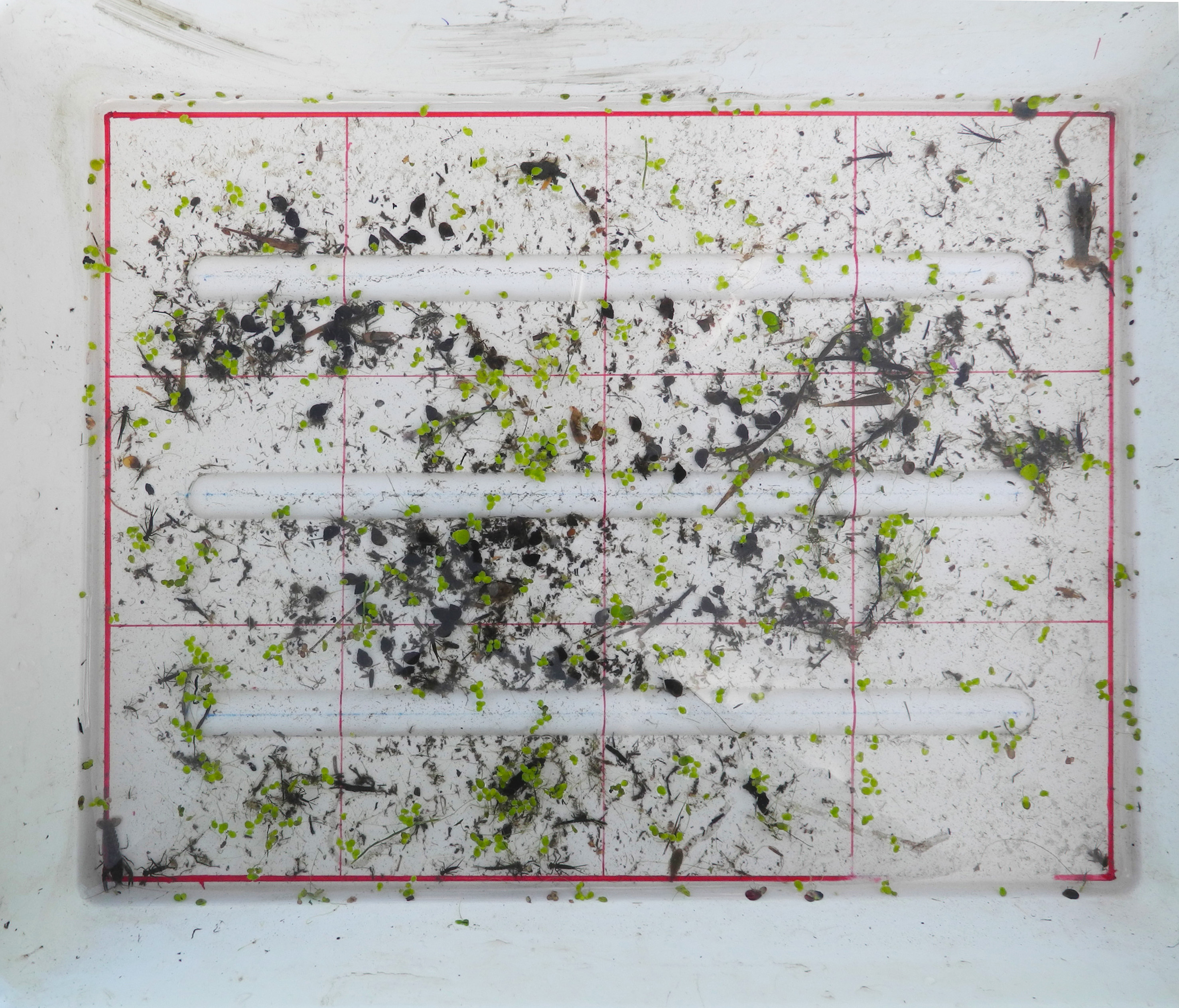
The result of a single sample digitalization is a pair of two dimensional images (Image 5 B and C, above). Those images must be proceeded using proper computer software (e.g. StereoPhoto Maker), in order to get three dimensional view. Afterwards taxa identification is carried out by 3D images analysis. Above described identification process differs from traditional one (using stereoscopic microscope). Therefore, it is recommended to use identification key developed for use in NoMBSI.Click this link to download free software for 3D images visualization and processing (StereoPhoto Maker).
 |
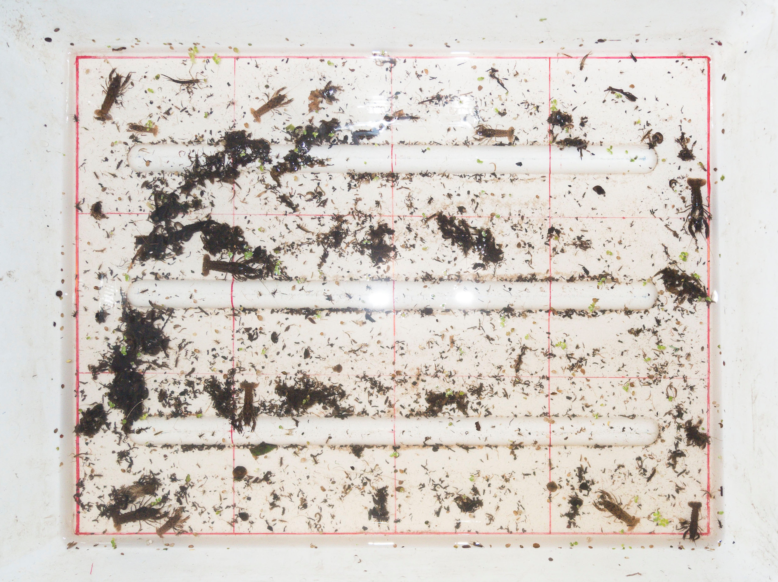 |
| River 1; upper left image, | upper right image |
 |
 |
| River 2; upper left image, | upper right image |
 |
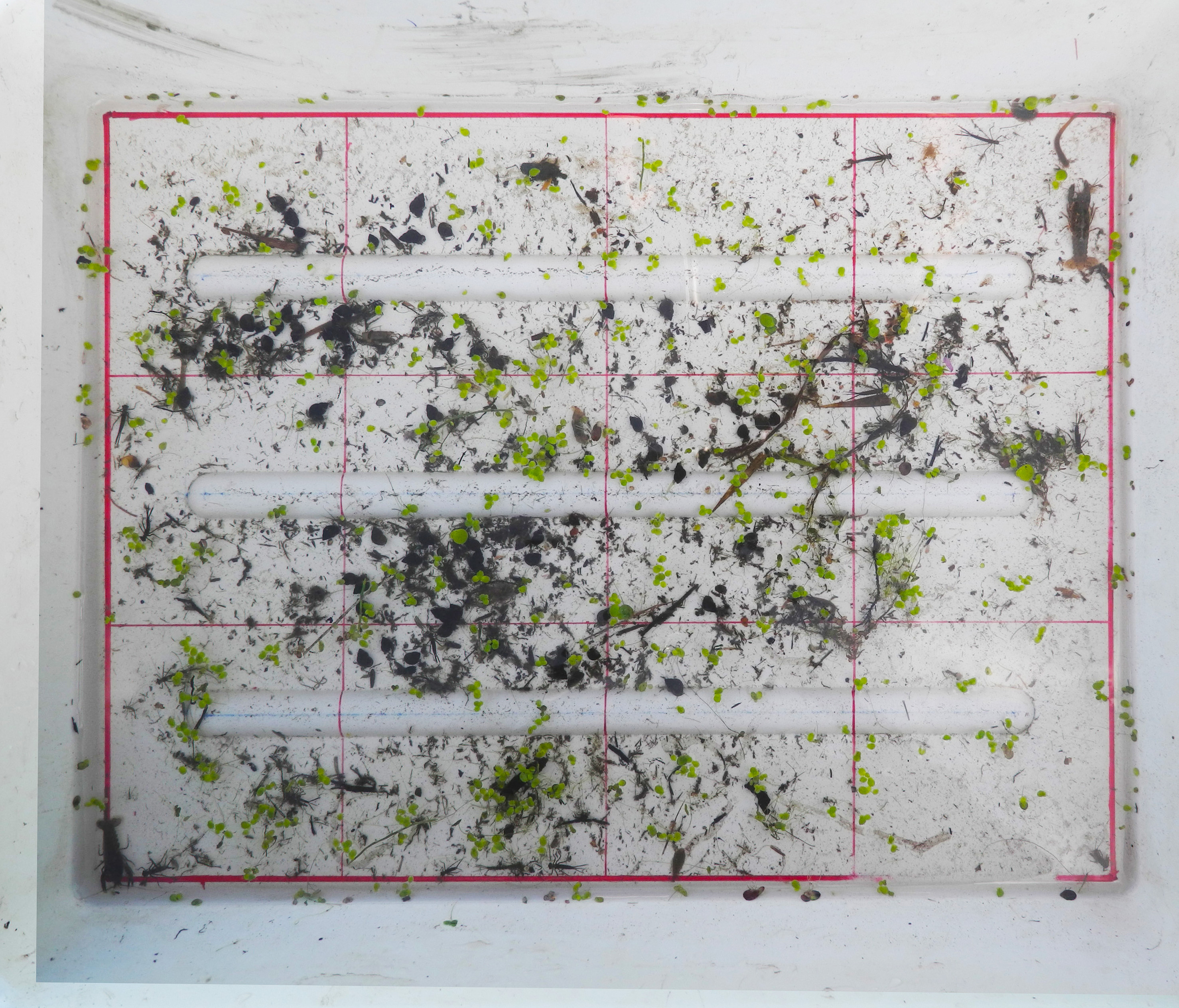 |
| River 3; upper left image, | upper right image |
 |
 |
| River 4; upper left image, | upper right image |
 |
 |
| River 5; upper left image, | upper right image |
(back to top of page)
Data interpretation – index xxx
Because NoMBSI is very selective and focused only on detection of a group of indicative taxa it is doubtful to reliably use indices currently employed in other assessment procedures. Therefore, it was necessary to develop a new metric (corresponding to the data provided by using NoMBSI).
Literature
- Czerniawska-Kusza I., 2004. Use of Artificial Substrates for Sampling Benthic Macroinvertebrates in the Assessment of Water Quality of Large Lowland Rivers. Polish Journal of Environmental Studies, 13: 579-584.
- De Pauw, N., D. Roels & A. P. Fontoura, 1986. Use of artificial substrates for standardized sampling of macroinvertebrates in the assessment of water quality by the Belgian Biotic Index. Hydrobiologia, 133: 237-258.
(back to top of page)